Groundwork for safe, predictable, and masterful application
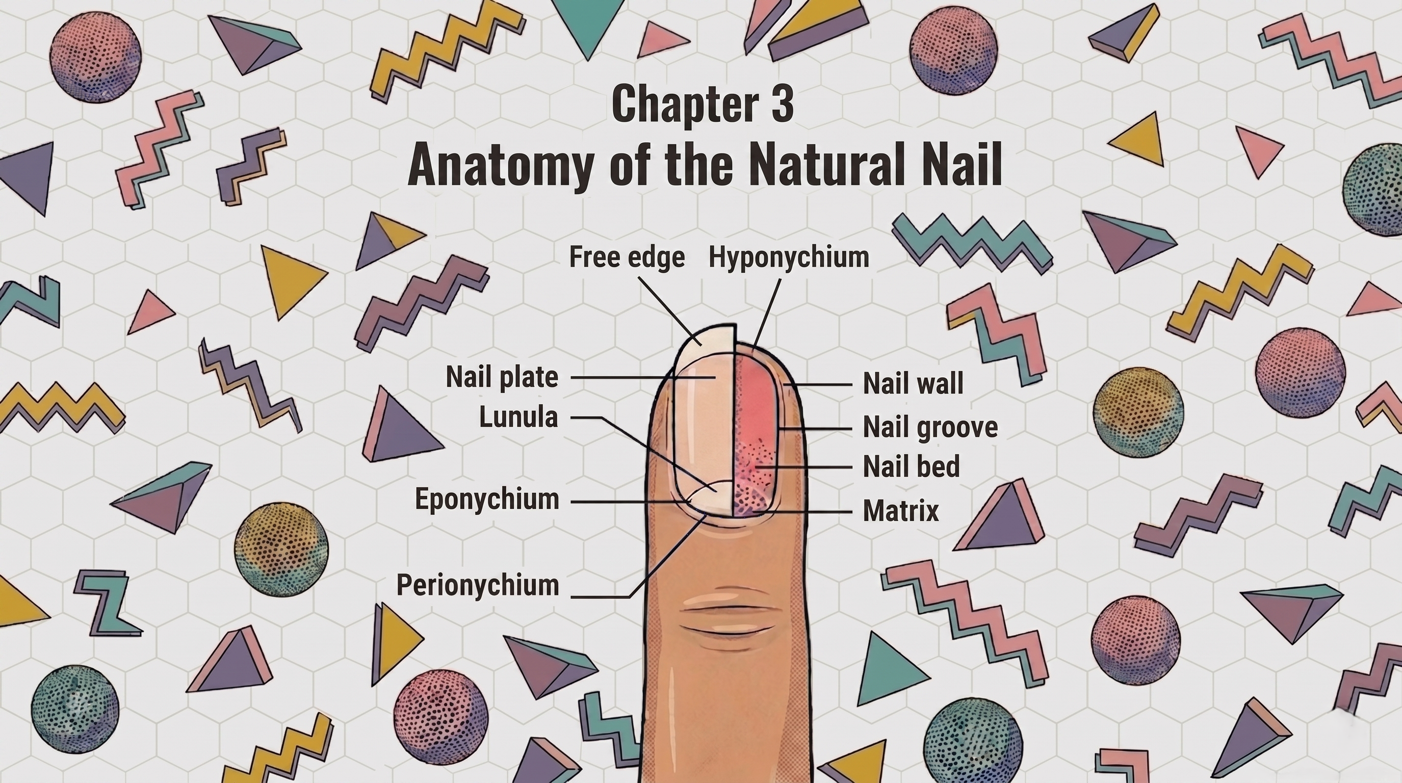
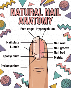
Chapter 3: Anatomy of the Natural Nail
Know Your Canvas
Before you can safely apply a single drop of acrylic chemistry, you must understand the anatomy you are working on. We often call the nail plate a “canvas,” but remember that it is a living extension of the client’s body. The natural nail plate is essentially a tough, fibrous protein called keratinized cellular tissue. Your goal is to strongly attach the artificial acrylic enhancement to this keratinized plate without damaging the structure underneath.
-
- The Matrix: This is the most vital biological area, hidden deep under the eponychium (the cuticle skin). It is where the natural nail cells are born and differentiated. The Matrix is responsible for the growth rate, thickness, and health of the natural nail. While you cannot see it, your application must never compress or damage this root, or permanent nail deformities will occur. ……….
- The Nail Plate: This is your durable, non-living keratin canvas. A hard layer that rests on the nail bed, it is thousands of overlapping cells thick. It is extremely receptive to adhesive chemistry, if prepared correctly.

- The Eponychium (is not the Cuticle): Warning for the technician: Acrylic will only form a permanent chemical bond with the keratin of the nail plate. It will never bond to living skin. This living, sensitive skin, often confused with the “cuticle” (dead tissue), must be carefully managed to prevent lifting, as any product left touching the skin creates a structural void where water and bacteria can enter.